ilustrações de stock, clip art, desenhos animados e ícones de digital illustration of signal travelling from spinal cord through brain stem, connecting nerve fibre with third-order neuron in thalamus to somatosensory cortex - spinal neuron
Representation Of The Transmission Of The Pain From Neuron To Neuron At The Level Of The Spinal Cord. The Presynaptic Neuron Releases The...
Photo Essay From Laboratory. Genetics Research Laboratory. Gene Therapy Requires A Gene Drug And A Vector To Carry It To The Target Cell. The Study...
Genetics Research Laboratory. Gene Therapy Requires A Gene Drug And A Vector To Carry It To The Target Cell. The Study Of These Vectors Vectorology...
The Path Of The Pain And Its Inhibition At The Level Of The Neurons Of The Spinal Cord. The Red Arrow Indicates The Path Of The Pain That Directs...
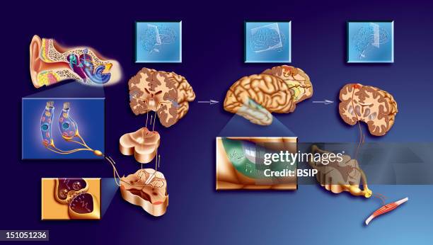
Sound Perception And Interpretation. When A Sound Reaches The Ear, It Arrives At The Cochlea Where Sense Receptors Send A Message To The Neurons. The...
Sensory pathways, motor pathways, sensory-motor pathways, reflex arc, stimulus, pain. The reflex arc is a neural circuit made up of a neuron...
Motor neuron, final pathways of the motor pathway system, innervation of muscle fibers. Motor neurons are cells that control the movement of the...
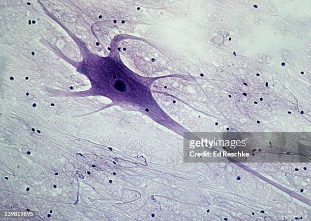
photomicrograph of spinal cord neuron of the grey matter showing cell body, nucleus and neucleolus; 250x. - spinal neuron imagens e fotografias de stock
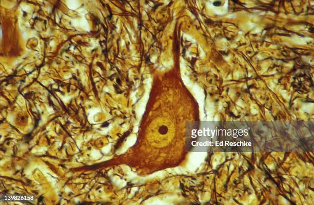
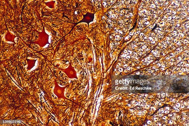
photomicrograph of motor neuron of spinal cord, showing cell body, nucleus, dendrites, axon, and neurolglia (black spots); 50x. - spinal neuron imagens e fotografias de stock
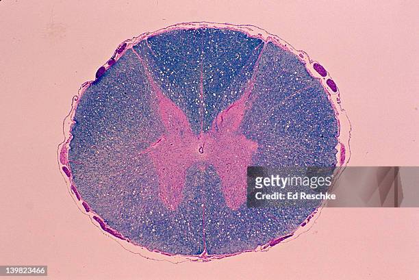
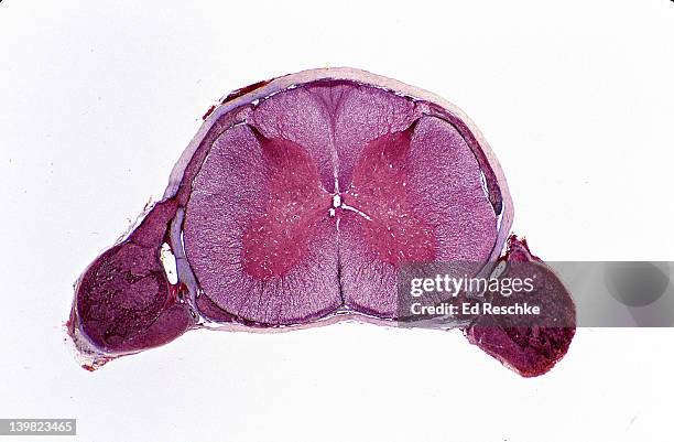
spinal cord. cross section, 5x shows: gray matter (inner pink, butterfly-shaped area), white matter (outer blue area), central canal, meninges, dorsal horn, lateral horn, ventral (anterior) horn, and anterior horn cells (motor neuron cell bodies). - spinal neuron imagens e fotografias de stock
spinal cord. cross section, 2.5x. shows: gray matter, white matter, dorsal root ganglia, dorsal and ventral roots, central canal, anterior horn cells (motor neuron cell bodies) & meninges - spinal neuron imagens e fotografias de stock
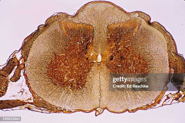
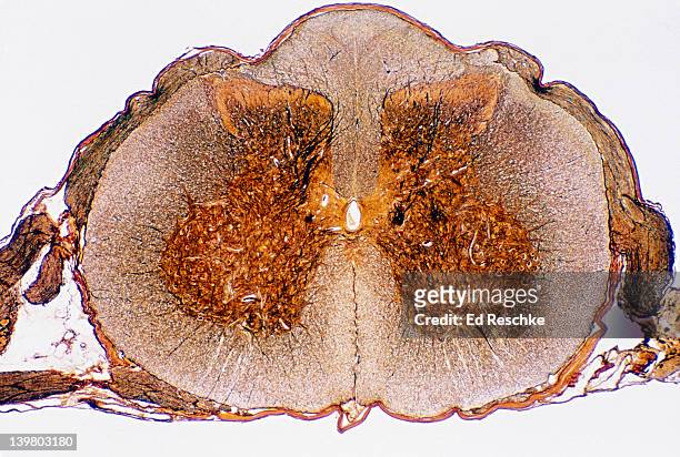
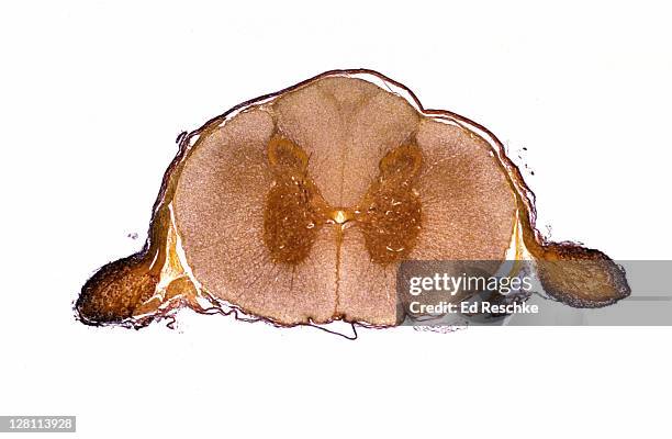
spinal cord cross section, 5x shows: gray matter (golden butterfly), white matter, central canal, dorsal & ventral root, dorsal root ganglion, meninges, dorsal horn, ventral (anterior) horn, & anterior horn cells (motor neuron cell bodies) - spinal neuron imagens e fotografias de stock
ganglion; dorsal root, spinal cord, sensory neuron cell bodies 100x at 35mm. shows: masses of sensory neuron cell bodies, nuclei, and nucleoli. - spinal neuron imagens e fotografias de stock
neuron (motor), spinal cord, 50x at 35mm. shows: cell body, nucleus, dendrites (many), and neuroglial cells (black dots). - spinal neuron imagens e fotografias de stock
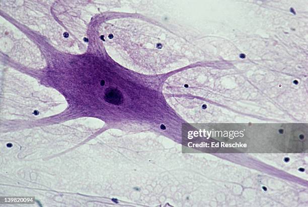
neuron (motor), spinal cord, 50x at 35mm. shows: cell body, nucleus, dendrites (several), axon (single, long nerve fiber), and neuroglial cells (black spots). - spinal neuron imagens e fotografias de stock
neuron (motor), spinal cord, 100x at 35mm. shows: cell body, nucleus, dendrites (several), axon (single, long nerve fiber), and neuroglial cells (black spots). - spinal neuron imagens e fotografias de stock
neuron (motor), spinal cord, 50x at 35mm. shows: cell body, nucleus, dendrites (several), axon (single, long nerve fiber), and neuroglial cells (black spots). - spinal neuron imagens e fotografias de stock
motor neuron; spinal cord, 50x at 35mm. shows: cell body, nucleus, dendrites (numerous processes attached to cell body), axon (single, long, nerve fiber), and neuroglial cells (dark spots). - spinal neuron imagens e fotografias de stock
motor neuron; spinal cord, 50x at 35mm. shows: cell body, nucleus, dendrites (numerous processes attached to cell body), axon (single, long, nerve fiber), and neuroglial cells (dark spots). - spinal neuron imagens e fotografias de stock
gray & white matter; spinal cord, 25x at 35mm. shows: gray matter (neuron cell bodies and unmyelinated nerve fibers), and white matter (myelinated nerve fibers in cross section). - spinal neuron imagens e fotografias de stock
dorsal root ganglion & dorsal root, spinal cord, 50x at 35mm. shows: the dorsal root ganglion (composed of masses of sensory neuron cell bodies with nuclei), and the dorsal root (composed of nerve fibers). - spinal neuron imagens e fotografias de stock
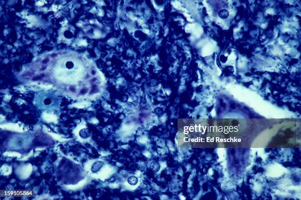
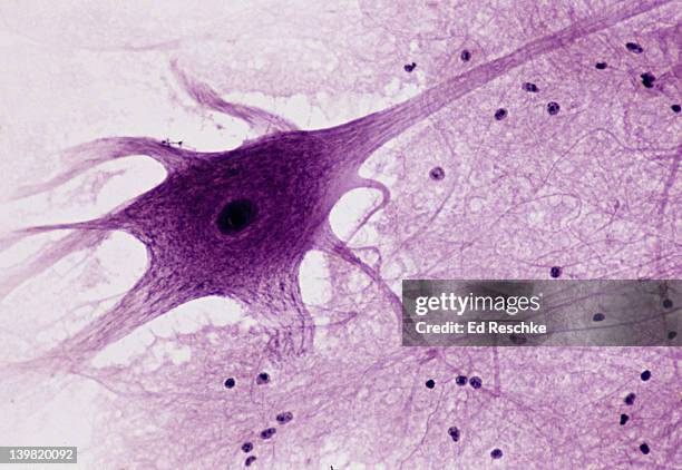
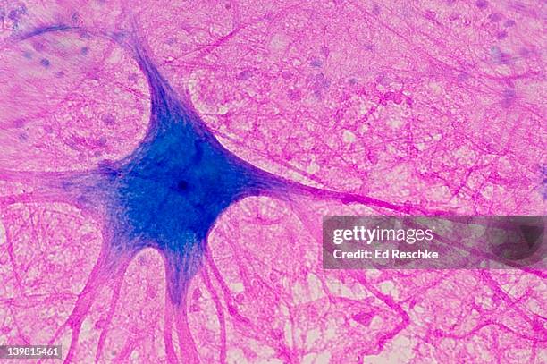
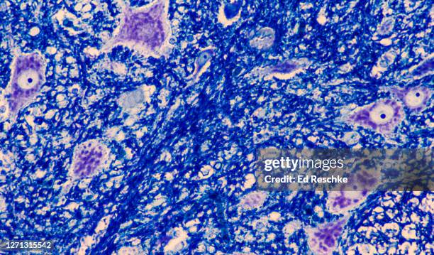
motor neuron (multipolar) with cell body and many processes (mostly dendrites), spinal cord (magnification x100). this multipolar motor neuron comes from the anterior (ventral) horn of the spinal cord grey matter. the stain is methylene blue and phloxine. - spinal neuron imagens e fotografias de stock
ganglion (dorsal root or spinal ganglion neurons). shows mass of sensory neuron cell bodies; 100x. cell bodies show nuclei and nucleoli - spinal neuron imagens e fotografias de stock
spinal cord, shows: neurons, grey matter with motor neuron cell bodies, white matter with myelinated nerve fibers. 50x - spinal neuron imagens e fotografias de stock
spinal cord, cross-section. shows motor neuron, dorsal and ventral horns. 5x - spinal neuron imagens e fotografias de stock
neuron cell bodies and unmyelinated nerve fibers from the gray matter of the spinal cord, 250x - spinal neuron imagens e fotografias de stock
spinal cord cross section. shows gray & white matter, central canal, motor neuron, dorsal and ventral roots. 3x - spinal neuron imagens e fotografias de stock
motor neuron cell bodies (anterior horn cells) with numerous nissl bodies (or chromatophilic substance), spinal cord, 100x - spinal neuron imagens e fotografias de stock
motor neuron (anterior horn cell) in the anterior horn of the spinal cord, gray matter, neuron cell body and unmyelinated nerve fibers, 250x - spinal neuron imagens e fotografias de stock
gray matter of the spinal cord--neuron cell bodies and unmyelinated nerve fibers, 50x - spinal neuron imagens e fotografias de stock
neuron cell bodies of sensory neurons in the dorsal (or posterior) root ganglion of the spinal cord, 100x - spinal neuron imagens e fotografias de stock
Site of action of morphine. Increased painful nerve impulses from the cutaneous, muscular, articular or visceral tissues from the nociceptors, in...
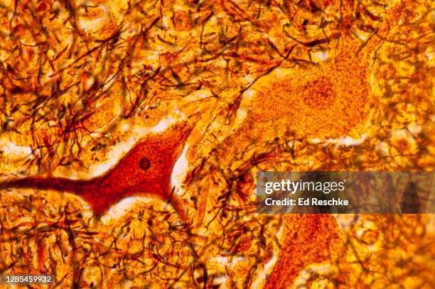
motor neuron--cell body, dendrites and axon, spinal cord, 100x - spinal neuron imagens e fotografias de stock